
Understanding ECG Changes in Acute Myocardial Infarction
Author: Dr A M Thirugnanam
Introduction
Acute myocardial infarction (AMI) remains one of the leading causes of death worldwide. Every minute of delay in diagnosis results in progressive myocardial damage, making rapid electrocardiographic interpretation one of the most valuable skills for physicians, emergency doctors, cardiologists, and medical students.
The 12-lead electrocardiogram (ECG) remains the fastest, most accessible, and most cost-effective investigation for diagnosing ST-elevation myocardial infarction (STEMI). Recognizing early ECG abnormalities enables immediate reperfusion therapy, significantly reducing mortality and preserving ventricular function.
This comprehensive guide explains ECG changes during acute myocardial infarction, their progression, localization, reciprocal changes, and clinical significance.
What Happens During Acute Myocardial Infarction?
Acute myocardial infarction occurs when coronary artery blood flow becomes abruptly interrupted due to plaque rupture and thrombus formation.
The ischemic myocardium undergoes three stages:
- Ischemia
- Injury
- Infarction
Each stage produces characteristic ECG findings that evolve over time.
Evolution of ECG Changes
ECG findings evolve in a predictable sequence.
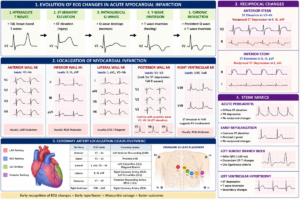
Stage 1: Hyperacute T Waves
The earliest ECG manifestation is the appearance of hyperacute T waves.
Characteristics include:
- Tall
- Broad-based
- Symmetrical
- Localized to affected leads
These changes may appear within minutes after coronary occlusion.
Clinical importance:
Hyperacute T waves often precede ST elevation and represent an opportunity for very early diagnosis.
Stage 2: ST-Segment Elevation
Persistent coronary occlusion produces myocardial injury resulting in ST-segment elevation.
Diagnostic criteria include:
- ≥1 mm elevation in two contiguous limb leads
- ≥2 mm elevation in contiguous precordial leads (depending on age and sex)
Typical appearance:
- Convex (“tombstone”)
- Straight
- Occasionally concave
Clinical importance:
Persistent ST elevation indicates complete coronary artery occlusion requiring urgent reperfusion.
Stage 3: Pathological Q Waves
Several hours after infarction, irreversible myocardial necrosis develops.
Characteristics include:
- Duration ≥40 ms
- Depth >25% of R wave
Not every STEMI develops pathological Q waves, especially after successful primary PCI.
Stage 4: T-Wave Inversion
As myocardial injury begins to heal, ST segments normalize while T waves become inverted.
These inverted T waves may persist for weeks or months.
Stage 5: Chronic Infarction
Late ECG findings include:
- Persistent pathological Q waves
- Loss of R-wave progression
- Normal ST segment
- Persistent T-wave inversion in some patients
Localization of Myocardial Infarction
The ECG identifies the affected coronary artery territory.
Anterior Wall Myocardial Infarction
Affected leads:
- V1
- V2
- V3
- V4
Usually caused by:
Left anterior descending artery occlusion.
Clinical features:
- Large infarct size
- Cardiogenic shock
- Heart failure
- High mortality
Septal Infarction
Leads involved:
- V1
- V2
Usually due to proximal LAD disease.
Lateral Wall Infarction
Leads:
- I
- aVL
- V5
- V6
Usually caused by:
- Left circumflex artery
- Diagonal branch
Inferior Wall Infarction
Leads:
- II
- III
- aVF
Usually caused by:
- Right coronary artery
- Left circumflex artery
Associated complications:
- Bradycardia
- AV block
- Right ventricular infarction
Posterior Myocardial Infarction
ECG findings:
- Tall R waves in V1-V3
- ST depression
- Upright T waves
Posterior leads:
- V7
- V8
- V9
Right Ventricular Infarction
Evaluate with:
Right-sided leads:
- V3R
- V4R
Typical findings:
ST elevation in V4R.
Reciprocal ECG Changes
Reciprocal changes increase diagnostic confidence.
Examples:
Anterior STEMI:
- ST elevation in V1-V4
- Reciprocal ST depression in II, III, aVF
Inferior STEMI:
- ST elevation in II, III, aVF
- Reciprocal depression in I and aVL
These findings strongly support acute coronary occlusion.
Coronary Artery Localization
| ECG Leads | Coronary Artery |
|---|---|
| V1-V4 | Left Anterior Descending |
| I, aVL | Diagonal or Left Circumflex |
| V5-V6 | Left Circumflex |
| II, III, aVF | Right Coronary Artery |
| V7-V9 | Posterior Descending Artery |
| V4R | Right Coronary Artery |
H2 STEMI Mimics
Several conditions resemble myocardial infarction.
Acute Pericarditis
Features:
- Diffuse ST elevation
- PR depression
- No reciprocal changes
Early Repolarization
Common in young adults.
Features:
- Concave ST elevation
- Stable ECG
- No reciprocal changes
Left Bundle Branch Block
Diagnosis requires modified Sgarbossa criteria.
Left Ventricular Hypertrophy
Produces secondary ST-T abnormalities that may mimic STEMI.
ECG Warning Signs Requiring Immediate Action
Urgent reperfusion should be considered when ECG demonstrates:
- New ST elevation
- Hyperacute T waves
- Posterior MI
- Right ventricular infarction
- New LBBB with ischemic symptoms
- De Winter pattern
- Wellens syndrome
Practical Approach to Reading an ECG in Suspected MI
Follow this systematic approach:
- Confirm patient symptoms.
- Assess rhythm.
- Measure heart rate.
- Determine electrical axis.
- Examine P waves.
- Measure PR interval.
- Assess QRS duration.
- Evaluate ST segment.
- Examine T waves.
- Identify pathological Q waves.
- Look for reciprocal changes.
- Localize infarction.
- Compare previous ECGs.
- Repeat ECG every 15–30 minutes if suspicion remains high.
Clinical Pearls
- A normal ECG does not exclude myocardial infarction.
- Repeat ECGs improve diagnostic accuracy.
- Hyperacute T waves may precede ST elevation.
- Posterior MI is frequently overlooked.
- Right ventricular infarction changes management significantly.
- Reciprocal changes strengthen STEMI diagnosis.
- Time to reperfusion remains the most important determinant of survival.
Conclusion
Rapid recognition of ECG changes in acute myocardial infarction is essential for saving lives. Understanding the sequential evolution from hyperacute T waves to ST elevation, pathological Q waves, and T-wave inversion enables clinicians to diagnose myocardial infarction early and initiate prompt reperfusion therapy.
Mastering ECG interpretation is an indispensable skill for every cardiologist, emergency physician, resident doctor, and medical student.
Frequently Asked Questions (FAQ)
What is the earliest ECG change in myocardial infarction?
Hyperacute T waves are usually the earliest ECG abnormality.
Which leads indicate anterior myocardial infarction?
V1 to V4 indicate anterior wall involvement.
What causes ST elevation?
Transmural myocardial injury secondary to complete coronary artery occlusion.
What are reciprocal changes?
ST depression occurring in leads opposite the infarcted territory.
Can myocardial infarction occur with a normal ECG?
Yes. Early ECGs may be normal. Serial ECGs and cardiac biomarkers are essential.
Internal Links
- Home → https://cardiologybooks.com/
- ECG Books → https://cardiologybooks.com/product-category/ecg/
- Cardiology eBooks → https://cardiologybooks.com/shop/
- Emergency Cardiology Books → https://cardiologybooks.com/product-category/emergency-cardiology/
- Free Cardiology Downloads → https://cardiologybooks.com/free-downloads/
- Blog → https://cardiologybooks.com/blog/
- About Dr A M Thirugnanam → https://cardiologybooks.com/about/